Builders and Blocks – Engineering Blood Vessels with Stem Cells

Back in 2001, when we first began studying how regenerative cells (stem cells or more mature progenitor cells) enhance blood vessel growth, our group as well as many of our colleagues focused on one specific type of blood vessel: arteries. Arteries are responsible for supplying oxygen to all organs and tissues of the body and arteries are more likely to develop gradual plaque build-up (atherosclerosis) than veins or networks of smaller blood vessels (capillaries). Once the amount of plaque in an artery reaches a critical threshold, the oxygenation of the supplied tissues and organs becomes compromised. In addition to this build-up of plaque and gradual decline of organ function, arterial plaques can rupture and cause  severe sudden damage such as a heart attack. The conventional approach to treating arterial blockages in the heart was to either perform an open-heart bypass surgery in which blocked arteries were manually bypassed or to place a tube-like “stent” in the blocked artery to restore the oxygen supply. The hope was that injections of regenerative cells would ultimately replace the invasive procedures because the stem cells would convert into blood vessel cells, form healthy new arteries and naturally bypass the blockages in the existing arteries.


Image of mouse red blood cells flowing through an engineered human blood vessel- Image from Paul and colleagues (2013), Creative Commons license.
Image of mouse red blood cells flowing through an engineered human blood vessel implanted in a mouse- Image from Paul and colleagues (2013), Creative Commons license.

As is often the case in biomedical research, this initial approach turned out to be fraught with difficulties. The early animal studies were quite promising and the injected cells appeared to stimulate the growth of blood vessels, but the first clinical trials were less successful. It was very difficult to retain the injected cells in the desired arteries or tissues, and even harder to track the fate of the cells. Which stem cells should be injected? Where should they be injected? How many? Can one obtain enough stem cells from an individual patient so that one could use his or her own cells for the cell therapy? How does one guide the injected cells to the correct location, and then guide the cells to form functional blood vessel structures? Would the stem cells of a patient with chronic diseases such as diabetes or high blood pressure be suitable for therapies, or would such a patient have to rely on stem cells from healthier individuals and thus risk the complication of immune rejection?

The complexity of blood-vessel generation became increasingly apparent, both when studying the biology of stem cells as well as when designing and conducting clinical trials. A large clinical study published in 2013 studied the impact of bone marrow cell injections in heart attack patients and concluded that these injections did not result in any sustained benefit for heart function. Other studies using injections of patients’ own stem cells into their hearts had led to mild improvements in heart function, but none of these clinical studies came close to fulfilling the expectations of cardiovascular patients, physicians and researchers. The upside to these failed expectations was that it forced the researchers in the field of cardiovascular regeneration to rethink their goals and approaches.

One major shift in my own field of interest – the generation of new blood vessels – was to reevaluate the validity of relying on injections of cells. How likely was it that millions of injected cells could organize themselves into functional blood vessels? Injections of cells were convenient for patients because they would not require the surgical implantation of blood vessels, but was this attempt to achieve a convenient therapy undermining its success? An increasing number of laboratories began studying the engineering of blood vessels in the lab by investigating the molecular cues which regulate the assembly of blood vessel networks, identifying molecular scaffolds which would retain stem cells and blood vessel cells and combining various regenerative cell types to build functional blood vessels. This second wave of regenerative vascular medicine is engineering blood vessels which will have to be surgically implanted into patients.  This means that it will be much harder to get approval to conduct such invasive implantations in patients than the straightforward injections which were conducted in the first wave of studies, but most of us who have now moved towards a blood vessel engineering approach feel that there is a greater likelihood of long-term success even if it may take a decade or longer till we obtain our first definitive clinical results.

The second conceptual shift which has occurred in this field is the realization that blood vessel engineering is not only important for treating patients with blockages in their arteries. In fact, blood vessel engineering is critical for all forms of tissue and organ engineering. In the US, more than 120,000 people are awaiting an organ transplant but only a quarter of them will receive an organ in any given year. The number of people in need of a transplant will continue to grow but the supply of organs is limited and many patients will unfortunately die while waiting for an organ which they desperately need. The advances in stem cell biology have made it possible to envision creating organs or organoids (functional smaller parts of an organ) which could help alleviate the need for organs. One thing that most organs and tissues need is a network of tiny blood vessels that permeate the whole tissue: small capillary networks. For example, a liver built out of liver cells could never function without a network of tiny blood vessels which supply the liver cells with metabolites and oxygen. From an organ engineering point of view, microvessel engineering is just as important as the building of functional arteries.

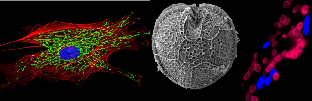
In one of our recent projects, we engineered functional human blood vessels by combining bone marrow derived stem cells with endothelial cells (the cells which coat the inside of all blood vessels). It turns out that stem cells do not become endothelial cells but instead release a molecular signal – the protein SLIT3- which instructs the endothelial cells to assemble into networks. Using a high resolution microscope, we watched this process in real-time over a course of 72 hours in the laboratory and could observe how the endothelial cells began lining up into tube-like structures in the presence of the bone marrow stem cells. The human endothelial cells were like building blocks, the human bone marrow stem cells were the builders “overseeing” the construction. When we implanted the assembled blood vessel structures into mice, we could see that they were fully functional, allowing mouse blood to travel through them without leaking or causing any other major problems (see image, taken from reference 3).

I am sure that SLIT3 is just one of many molecular cues released by the stem cells to assemble functional networks and there are many additional mechanisms which still need to be discovered. We still need to learn much more about which “builders” and which “building blocks” are best suited for each type of blood vessel that we want to construct. The fact that human fat tissue can serve as an important resource for obtaining adult stem cells(“builders”) is quite encouraging, but we still know very little about the overall longevity of the engineered vessels, the best way to implant them into patients, and the key molecular and biomechanical mechanisms which will be required to engineer organs with functional blood vessels. It will be quite some time until the first fully engineered organs will be implanted in humans, but the dizzying rate of progress suggests that we can be quite optimistic.

(An earlier version of this article was first published on 3Quarksdaily.com).


References and links (all of them are open access, so you can read them for free, including the original paper we published):

 1. A recent longform overview article I wrote for “The Scientist” in which I describe the importance of blood vessel engineering for organ engineering:

J Rehman “Building Flesh and Blood“, The Scientist (2014), 28(5):48-53


2. An unusual and abundant source of adult stem cells which promote the formation of blood vessels: Fat tissue obtained from individuals who undergo a liposuction! 

J Rehman “The Power of Fat” Aeon Magazine (2014)


3. The study which describes how adult stem cells release a protein (SLIT3) which organizes blood vessel cells into functional networks (open access – can be read free of charge):

J.D. Paul et al., “SLIT3-ROBO4 activation promotes vascular network formation in human engineered tissue and angiogenesis in vivo” J Mol Cell Cardiol (2013), 64:124-31.


ResearchBlogging.org

Paul JD, Coulombe KL, Toth PT, Zhang Y, Marsboom G, Bindokas VP, Smith DW, Murry CE, & Rehman J (2013). SLIT3-ROBO4 activation promotes vascular network formation in human engineered tissue and angiogenesis in vivo. Journal of molecular and cellular cardiology, 64, 124-31 PMID: 24090675

Does Human Fat Contain Stem Cells?

Aeon Magazine recently published my longform essay on our research with human liposuction samples and our attempts to use fat for regenerative and therapeutic purposes. Many research groups, including our own group, have been able to isolate stem cells from human fat. However, when it came to using this cells for treating cardiovascular disease, the cells behaved in a manner that we had not anticipated.

Undifferentiated mesenchymal stem cells (left) and their fat neighbors (right)
Undifferentiated mesenchymal stem cells (left) and their fat neighbors (right) – From our PLOS One paper

We were unable to convert them into heart muscle cells or blood vessel endothelial cells, but we found that they could help build large networks of blood vessels by releasing important growth factors. Within a few years of our initial publication, clinical trials with patients with blocked arteries or legs were already being planned, and are currently underway.

We decided to call the cells “adipose stromal cells” because we wanted to emphasize that they were acting as a “stroma” (i.e. supportive environment for blood vessels) and not necessarily as stem cells (i.e. cells that convert from an undifferentiated state into mature cell types). In other contexts, these same cells were indeed able to act like “stem cells”, because they could be converted into bone-forming or cartilage-forming cells, thus showing the enormous versatility and value of the cells that reside within our fat tissues.

The answer to the question “Does Human Fat Contain Stem Cells?” is Yes, but these cells cannot be converted into all desired tissues. Instead, they have important supportive functions that can be used to engineer new blood vessels, which is a critical step in organ engineering.

In addition to describing our scientific work, the essay also mentions the vagaries of research, the frustrations I had as a postdoctoral fellow when my results were not turning out as I had expected, and how some predatory private clinics are already marketing “fat-derived stem cell therapies” to paying customers, even though the clinical results are still rather preliminary.


For the readers who want to dig a bit deeper, here are some references and links:


1. The original paper by Patricia Zuk and colleagues which described the presence of stem cells in human liposuction fat:

Zuk, P et al (2001) “Multilineage Cells from Human Adipose Tissue: Implications for Cell-Based Therapies


2. Our work on how the cells can help grow blood vessels by releasing proteins:

Rehman, J et al (2004) “Secretion of Angiogenic and Antiapoptotic Factors by Human Adipose Stromal Cells


3. Preliminary findings from ongoing clinical studies in which heart attack patients receive infusions of fat derived cells into their hearts to improve heart function and blood flow to the heart:

Houtgraf, J et al (2012) “First Experience in Humans Using Adipose Tissue–Derived Regenerative Cells in the Treatment of Patients With ST-Segment Elevation Myocardial Infarction


4. Preliminary results from an ongoing trial using the fat-derived cells in patients with severe blockages of leg arteries:

Bura, A et al (2014) “Phase I trial: the use of autologous cultured adipose-derived stroma/stem cells to treat patients with non-revascularizable critical limb ischemia


5. Example of how “cell therapies” (in this case bone marrow cells) are sometimes marketed as “stem cells” but hardly contain any stem cells:

The Largest Cell Therapy Trial in Heart Attack Patients Uses Hardly Any Stem Cells


6. The major scientific society devoted to studying the science of fat and its cells as novel therapies is called International Federation for Adipose Therapeutics and Science (IFATS).

I am not kidding, it is I-FATS!

Explore their website if you want to learn about all the exciting new research with fat derived cells.


7. Some of our newer work on how bone marrow mesenchymal stem cells turn into fat cells and what role their metabolism plays during this process:

Zhang, Y et al (2013) “Mitochondrial Respiration Regulates Adipogenic Differentiation of Human Mesenchymal Stem Cells


ResearchBlogging.org

Zuk PA, Zhu M, Mizuno H, Huang J, Futrell JW, Katz AJ, Benhaim P, Lorenz HP, & Hedrick MH (2001). Multilineage cells from human adipose tissue: implications for cell-based therapies. Tissue engineering, 7 (2), 211-28 PMID: 11304456


ResearchBlogging.org
Rehman J, Traktuev D, Li J, Merfeld-Clauss S, Temm-Grove CJ, Bovenkerk JE, Pell CL, Johnstone BH, Considine RV, & March KL (2004). Secretion of angiogenic and antiapoptotic factors by human adipose stromal cells. Circulation, 109 (10), 1292-8 PMID: 14993122

Stem Cells and Their Fat Neighbors

We recently published a PLOS ONE paper (Mitochondrial respiration regulates adipogenic differentiation of human mesenchymal stem cells) in which we studied how the metabolism of an adult stem cell can influence its ability to differentiate. Human bone marrow mesenchymal stem cells (also known as marrow stromal cells, marrow progenitor cells or MSCs) can be converted into fat (adipocytes), cartilage (chondrocytes) or bone (osteoblasts). The work performed by Yanmin Zhang and Glenn Marsboom in my lab showed that MSCs undergo a major metabolic shift towards increased mitochondrial oxidation when they become fat cells and that suppressing mitochondrial respiration can prevent their differentiation. The metabolic state of the adult stem cells is therefore not only an indicator of their “stemness”, it can be used to either promote or suppress their differentiation.


Dr. Peter Toth, one of the co-authors on the paper, helped us acquire some really beautiful images of the cells that I would like to share with the readers of the blog. The image below shows undifferentiated adult human bone marrow mesenchymal stem cells (MSCs) that were exposed to an adipogenic differentiation medium, i.e a combination of factors which induces the formation of fat cells (adipocytes). However, as with many stem cell differentiation protocols, not all stem cells turned into fat cells. The cells on the right have a typical fat-like structure in which cells are full of round lipid droplets. The neighboring cells on the left are MSCs that have not (yet?) become fat cells. We stained the cells with the fluorescent mitochondrial dye JC-1. Depolarized mitochondria appear green and hyperpolarized mitochondria red. As you can see, the cells on the left have a much higher mitochondrial membrane potential (significant amount of red among the green mitochondria) than their fat neighbors on the right (mostly green mitochondria, all of them located between lipid droplets). By capturing both cell types next to each other, we could show an illustrative example of how entwined metabolism and stem cell differentiation are. The morphology and metabolic state of neighboring cells in this image were quite different, despite the fact that all cells were subjected to the same cocktail of differentiation factors. The blue-appearing dye is DAPI and stains nuclei of cells so one can tell the cells apart. Each cell in this image has one blue nucleus.


The image was published with a PLoS ONE CC-BY license. Feel free to use it as an example of adult stem cell differentiation or how mitochondrial morphology and function can vary between stem cell and its differentiated progeny, as long as you attribute the original PLoS One paper. The image in the paper also has a scale bar and asterisks/arrows pointing out the specific cells.


ResearchBlogging.org

Zhang Y, Marsboom G, Toth PT, & Rehman J (2013). Mitochondrial respiration regulates adipogenic differentiation of human mesenchymal stem cells. PLoS ONE 8(10): e77077;  PMID: 24204740; DOI: 10.1371/journal.pone.0077077

Replicability of High-Impact Papers in Stem Cell Research

I recently used the Web of Science database to generate a list of the most highly cited papers in stem cell research. As of July 2013, the search for original research articles which use the key word “stem cells” resulted in the following list of the ten most widely cited papers to date:

Human ES cell colony – Nuclei labeled in blue, Mitochondria labeled in green- Rehman lab.1. Pittenger M et al. (1999) Multilineage potential of adult human mesenchymal stem cells. Science 284(5411):143-147

Citations: 8,157

2.  Thomson JA et al. (1998) Embryonic stem cell lines derived from human blastocysts. Science 282(5391):1145-1147

Citations: 5,565

3. Takahashi K and Yamanaka S (2006) Induction of pluripotent stem cells from mouse embryonic and adult fibroblast cultures by defined factors. Cell 126(4): 663-676

Citations: 5,034

4. Takahashi K et al. (2007) Induction of pluripotent stem cells from adult human fibroblasts by defined factors. Cell 131(5):861-872

Citations: 4,061

5. Donehower LA et al  (1992) Mice deficient for p53 are developmentally normal but susceptible to spontaneous tumours. Nature 356(6366): 215-221

Citations: 3,279

6. Al-Hajj M et al (2003) Prospective identification of tumorigenic breast cancer cells. Proceedings of the National Academy of Sciences 100(7): 3983-3988

Citations: 3,183

 7. Yu J et al (2007) Induced pluripotent stem cell lines derived from human somatic cells. Science 318(5858): 1917-1920

Citations: 3,086

 8. Jiang YH et al (2002) Pluripotency of mesenchymal stem cells derived from adult marrow. Nature 418(6893):41-49

Citations: 2,983

9. Orlic D et al (2001) Bone marrow cells regenerate infarcted myocardium. Nature 410 (6829):701-705

Citations: 2,961

10. Lu J et al (2005) MicroRNA expression profiles classify human cancers. Nature 435(7043): 834-838

Citations: 2,917


Three of the articles (Donehower et al, Al-Hajj et al and Lu et al) in this “top ten list” do not focus on stem cells but are actually cancer research papers. They were probably identified by the search because the authors may have made comparisons to stem cells or used stem cells as tools.The remaining seven articles are indeed widely known in the stem cell field.

The Science paper by Pittenger and colleagues in 1999 provided a very comprehensive description of mesenchymal stem cells (MSCs), a type of adult stem cell which is found in the bone marrow alongside hematopoietic stem cells (HSCs). Despite the fact that MSCs and HSCs are both adult stem cells in the bone marrow, they have very different functions. HSCs give rise to circulating blood cells, whereas MSCs primarily form bone, fat and cartilage as was nicely demonstrated by Pittenger and colleagues.

The article by Thomson and colleagues was published in 1998 in the journal Science described the derivation of human embryonic stem cells (ESCs) and revolutionized the field of stem cell research. While adult stem cells have a very limited capacity in terms of lineages they can turn into, ESCs are derived from the early blastocyst stage of embryonic development (within the first 1-2 weeks following fertilization) and thus retain the capacity to turn into a very wide range of tissues, such as neurons, heart cells, blood vessel cells or liver cells. This paper not only identified the methods for isolating human ESCs, but also how to keep them in culture and expand them as undifferentiated stem cells.

The Cell paper by Takahashi and Yamanaka in 2006 represented another major advancement in the field of stem cell biology, because it showed for the first time that a mouse adult skin cell (fibroblast) could be reprogrammed and converted into a truly pluripotent stem cell (an induced pluripotent stem cell or iPSC) which exhibited all the major characteristics of an embryonic stem cell (ESC). It was as if the adult skin cell was traveling back in time, erasing its identity of having been a skin cell and returning to primordial, embryonic-like stem cell. Only one year later, Dr. Yamanaka’s group was able to demonstrate the same phenomena for adult human skin cells in the 2007 Cell paper (Takahashi et al), and in the same year a different group independently confirmed that adult human cells could be reprogrammed to the iPSC state (Science paper by Yu et al in 2007). The generation of iPSCs described in these three papers is probably the most remarkable discovery in stem cell biology during the past decade. It is no wonder that each of these three papers have been cited several thousand times even though they were published only six or seven years ago, and that Dr. Yamanaka was awarded the 2012 Nobel prize for this pioneering work.

All five of the above-mentioned stem cell papers have one thing in common: the results have been repeated and confirmed by numerous independent laboratories all over the world. However, this does not necessarily hold true for the other two highly cited stem cell papers on this list.

The 2002 Nature paper by Jiang and colleagues from Dr. Verfaillie’s laboratory at the University of Minnesota proposed that the bone marrow contained a rather special subset of adult MSCs which had a much broader differentiation potential than had been previously recognized. While adult MSCs were thought to primarily turn into bone, cartilage or fat when given the appropriate cues, this rare new cell type – referred to as MAPCs (multipotent adult progenitor cells) – appeared to differentiate into a much broader range of tissues. The paper even showed data from an experiment in which these adult mouse bone marrow stem cells were combined with embryonic cells and gave rise to a chimeric mouse. i.e. a mouse in which the tissues were in part derived from standard embryonic cells and in part from the newly discovered adult MAPCs. Such chimerism suggested that the MAPCs were embryonic-like, contributing to the formation of all the tissues in the mice. At the time of its publication, this paper was met with great enthusiasm because it proved that the adult body contained embryonic-like cells, hidden away in the bone marrow, and that these MAPCs could be used to regenerate ailing organs and tissues without having to use ethically problematic human embryonic stem cells.

There was just one major catch. Many laboratories around the world tried to replicate the results and were unable to identify the MAPCs, and even when they found cells that were MAPCs, they were unable to confirm the embryonic-like nature of the cells. In a remarkable example of investigative journalism, the science journalists Peter Aldhous and Eugenie Reich identified multiple irregularities in the publications involving MAPCs and documented the inability of researchers to replicate the findings by publishing the results of their investigation in the New Scientist (PDF).

The second high profile stem cell paper which was also plagued by an inability to replicate the results was the 2001 Nature paper by Orlic and colleagues. In this paper from Dr. Anversa’s laboratory, the authors suggested that adult hematopoietic (blood-forming) stem cells from the bone marrow could regenerate an infarcted heart by becoming heart cells (cardiomyocytes). It was a rather bold claim, because simply injecting these blood-forming stem cells into the heart seemed to be sufficient to redirect their fate. Instead of giving rise to red and white blood cells, these bone marrow cells were generating functional heart cells. If this were the case, then every patient could be potentially treated with their own bone marrow and grow back damaged heart tissue after a heart attack. Unfortunately, it was too good to be true. Two leading stem cell laboratories partnered up to confirm the results, but even after years of experiments, they were unable to find any evidence of adult bone marrow stem cells converting into functional heart cells. They published their findings three years later, also in the journal Nature:

Murry CE et al (2004) Haematopoietic stem cells do not transdifferentiate into cardiac myocytes in myocardial infarcts. Nature 428(6983): 664-668

Citations: 1,150

Interestingly, the original paper which had made the claim that bone marrow cells can become functional heart cells has been cited nearly 3,000 times, whereas the refutation by Murry and colleagues, published in the same high-profile journal has been cited only 1,150 times. The vast majority of the nearly 3,000 citations of the 2001 paper by Orlic and colleagues occurred after it had been refuted in 2004! The 2001 Orlic et al paper has even been used to justify clinical trials in which bone marrow was obtained from heart attack patients and injected into their hearts. As expected after the refutation by Murry and colleagues, the success of these clinical trials was rather limited One of the largest bone marrow infusion trials in heart attack patients was recently published, showing no success of the therapy.

These claims of the two papers (Orlic et al and Jiang et al) were quite innovative and exciting, and they were also published in a high-profile, peer-reviewed journal, just like the other five stem cell papers. The crucial difference was the fact that their findings could not be replicated by other laboratories. Despite their lack of replicability, both papers had an enormous impact on the field of stem cell research. Senior scientists, postdocs and graduate students may have devoted a substantial amount of time and resources to developing projects that built on the findings of these two papers, only to find out that they could not be replicated. If there is a lesson to be learned, it is that we need to be rather cautious in terms of our enthusiasm for new claims in stem cell biology until they have been appropriately confirmed by other researchers. Furthermore, we need to streamline the replicability testing process so that we do not have to wait years before we find out that one of the most highly prized discoveries cannot be independently confirmed.


Update 7/24/2013: Peter Aldhous reminded me that the superb job of investigative journalism into the question of MAPCs was performed in partnership with the science writer Eugenie Reich, the author of a book on scientific fraud. I have updated the blog post to reflect this.

Stemming the Flow: Using Stem Cells To Treat Urinary Bladder Dysfunction

Neurogenic bladder is a disorder which occurs in spinal cord diseases such as spina bifida and is characterized by an inability of the nervous system to properly control the urinary bladder and the muscle tissue contained in the bladder wall. This can lead to spasms and a build-up of pressure in the bladder, often resulting in urinary incontinence. Children with spina bifida and neurogenic bladder often feel urges to urinate after drinking comparatively small amounts of liquid and they can also involuntarily leak urine. This is a source of a lot of emotional stress, especially in social settings such as when they are around friends or in school. If untreated, the long-standing and frequent pressure build-up in the bladder can have even more devastating effects such as infections or kidney damage.

Current treatments for neurogenic bladder involve surgeries which reconstruct and increase the size of the bladder by using tissue patches obtained from the bowel of the patient. Since such a gastrointestinal patch is derived from the patient’s own body, it is less likely to elicit an immune response and these intestinal tissue patches tend to be strong enough to withstand the pressures in the bladder. Unfortunately, the incompatibility of intestinal tissue and bladder tissue can lead to long-term complications, such as urinary tract infections, formation of urinary tract stones and in some rare cases even cancers. For this reason, researchers have been searching for newer safer patches which resemble the actual bladder wall.


A team of researchers at Northwestern University recently published a study which used stem cells of children with spina bifida to generate tissue patches that could be used for bladder surgery. In the paper “Cotransplantation with specific populations of spina bifida bone marrow stem/progenitor cells enhances urinary bladder regeneration” published in the Proceedings of the National Academy of Sciences (online publication on February 19, 2013), Arun Sharma and colleagues isolated two types of cells from the bone marrow of children with spina bifida: Mesenchymal stem cells (MSCs) and CD34+ cells (stem and progenitor cells which usually give rise to blood cells). They then coated a special polymer scaffold called POC with the cells and implanted this newly created patch into a rat bladder after performing a bladder augmentation surgery, similar to what is performed in patients with spina bifida. They then assessed the survival and formation of human muscle tissue on the implanted patch. When both human cell types (MSCs and CD34+) were combined, more than half of the implanted patch was covered with muscle tissue, four weeks after the implantation. If they only used CD34+ cells, they found that only a quarter of the patch was covered with muscle tissue. What is even more remarkable is that in addition to the newly formed muscle tissue, the implanted patch also showed evidence of some peripheral nerve growth and of blood vessel formation, both of which are found in healthy, normal bladder walls. These findings suggest that a patient’s own bone marrow stem cells can be used to help construct a tissue patch which could be used for bladder augmentation surgeries. The observation of some nerve growth in the implanted patch is also an exciting finding. One could conceivably try to re-connect the reconstructed bladder tissue with the main nervous system, but its success would largely depend on the severity of the neurologic disease.

One has to keep in mind that there are some key limitations to this study. The authors of the paper believe that the newly formed muscle tissue on the implanted patches was all derived from the patients’ bone marrow stem cells. However, there were no experiments performed to convincingly demonstrate this. The authors report that in previous studies, merely implanting the empty POC scaffold without any human stem cells resulted in 20% coverage with muscle tissue. This suggests that a big chunk of the newly formed muscle tissue is actually derived from the host rat and not from human stem cells. The authors also did not compare the effectiveness of this newly formed stem cell patch to the currently used intestinal patches, and there is no assessment of whether the newly formed muscle tissue on the reconstructed bladder is less prone to spasms and involuntary contractions. Lastly, all the in vivo testing of the tissue patches was performed in rats without neurogenic bladder and it is possible that the highly successful formation of muscle tissue may have been diminished if the animals had a neurologic disease.

A second study published in PLOS One took a different approach. In “Evaluation of Silk Biomaterials in Combination with Extracellular Matrix Coatings for Bladder Tissue Engineering with Primary and Pluripotent Cells” (online publication February 7, 2013), Debra Franck and colleagues describe how they coated a scaffold consisting of silk threads with extracellular matrix proteins such as fibronectin. Instead of using bone marrow stem cells, they converted induced pluripotent stem cells into the smooth muscle cells that are typically found inside the bladder wall and placed these newly differentiated cells on the silk scaffold. The induced pluripotent stem cells (iPSCs) used by Franck and colleagues can be generated from a patient’s own skin cells which reduces the risk of being rejected by a patient’s immune system. The advantage of this approach is that it starts out with a pure and truly pluripotent stem cell population, which is easier to direct and control than bone marrow stem cells. There are also a few important limitations to this second study. Franck and colleagues used mouse pluripotent stem cells and it is not clear that their approach would necessarily work with human pluripotent stem cells. They also did not test the function of these differentiated cells on the silk scaffold to check if they actually behaved like true bladder wall smooth muscle cells. Unlike the first study, Franck and colleagues did not evaluate the newly created patch in an animal model.

Both studies are purely experimental and much additional work is needed before they can be tested in humans, but both show promising new approaches to help improve bladder dysfunction. It is heartening to see that researchers are developing new cell-based therapies to help children and adults who suffer from neurogenic bladder. The results from these two experimental studies are still too preliminary to predict whether cell-based therapies can be successfully used in patients, but they represent important first steps.


Image credit: Taken from Franck D, Gil ES, Adam RM, Kaplan DL, Chung YG, et al. (2013) Evaluation of Silk Biomaterials in Combination with Extracellular Matrix Coatings for Bladder Tissue Engineering with Primary and Pluripotent Cells. PLoS ONE 8(2): e56237. doi:10.1371/journal.pone.0056237- Figure 6 B: Differentiated mouse induced pluripotent stem cells cultured on fibronectin-coated silk matrices show protein markers typically found in bladder smooth muscle cells.

ResearchBlogging.org
Franck, D., Gil, E., Adam, R., Kaplan, D., Chung, Y., Estrada, C., & Mauney, J. (2013). Evaluation of Silk Biomaterials in Combination with Extracellular Matrix Coatings for Bladder Tissue Engineering with Primary and Pluripotent Cells PLoS ONE, 8 (2) DOI: 10.1371/journal.pone.0056237